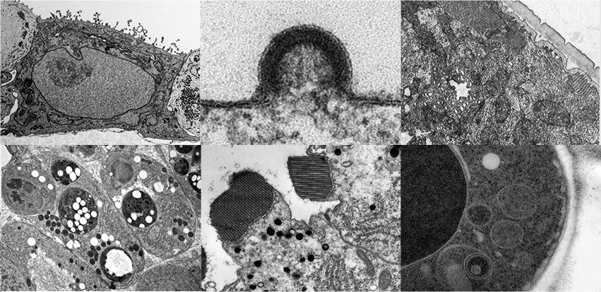
Electron microscopy (EM) provides a unique view of biological systems, allowing the visualization of the ultrastructure of your sample with nm resolution. It has provided the basis of our understanding of tissue and cellular architecture and continues to reveal a wealth of information about how cells and tissues function in health and disease.
The LMCB has been specialising in cell biological EM for over 20 years. The facility comprises state-of-the-art specimen preparation and imaging technologies which, together with two experienced researchers, support the needs of the LMCB groups in all their EM research.
With experience of a wide range of electron microscopy techniques as well as a broad range of model organisms, we can provide advice, training and practical support for researchers throughout the entire timeline of their projects. For example: planning experiments, sample preparation, imaging and data analysis. The support given is tailored to each project, and can vary from advice and technical support for the more experienced, full training in required techniques and the equipment use for the less experienced researchers or the undertaking of the EM projects ourselves, a collaborative endeavour as part of the ongoing research.
 Close
Close


